Home » Without Label » Leg Muscle Diagram : Anatomy Leg Muscles By Quarter Virus On Deviantart / The muscles that make up the quadriceps are the strongest and leanest of all muscles in the body.
Leg Muscle Diagram : Anatomy Leg Muscles By Quarter Virus On Deviantart / The muscles that make up the quadriceps are the strongest and leanest of all muscles in the body.
Leg Muscle Diagram : Anatomy Leg Muscles By Quarter Virus On Deviantart / The muscles that make up the quadriceps are the strongest and leanest of all muscles in the body.. These muscles include the gluteus maximus muscle (the largest muscle in the body) and the hamstrings group, which consists of the biceps femoris, semimembranosus, and semitendinosus muscles. This is important to understand the actions of the thigh muscles in limb movement. The aim of this exercise is to improve your confidence in identifying different structures. Notice the upper leg has a biceps muscle just like the upper arm does. The muscles that make up the quadriceps are the strongest and leanest of all muscles in the body.
Muscle and bone anatomy 12 photos of the muscle and bone anatomy back muscles and bones anatomy, human muscle and bone anatomy, muscle & bone anatomy 3d free download, muscle and bone anatomy app, muscle and bone anatomy quiz, human muscles, back muscles and bones anatomy, human muscle and bone anatomy, muscle & bone. The largest muscle masses in the leg are present in the thigh and the calf. Spend some time revising this diagram by connecting the name and location of each structure with what you've just learned in the video. See more ideas about muscle anatomy, human anatomy and physiology, body anatomy. The muscles that make up the quadriceps are the strongest and leanest of all muscles in the body.
Anatomy Of The Quadriceps Muscles from www.verywellfit.com The legs include the upper leg, knee, lower leg, ankle, and. The gastrocnemius muscle has two large bellies, called the medial head and the lateral head, and inserts into the calcaneus bone of the foot via its calcaneal tendon (also known as the achilles tendon.) Muscle and bone anatomy 12 photos of the muscle and bone anatomy back muscles and bones anatomy, human muscle and bone anatomy, muscle & bone anatomy 3d free download, muscle and bone anatomy app, muscle and bone anatomy quiz, human muscles, back muscles and bones anatomy, human muscle and bone anatomy, muscle & bone. The biceps femoris is a muscle of the posterior thigh composed of a long head and a short head. The 3 muscles are called triceps coxae. The calf muscle, on the back of the lower leg, is actually made up of two muscles: The muscles work together to enable movement and keep the hip in alignment. Observe the leg muscle diagram posted above and notice that there are many parts in the muscles.
Spend some time revising this diagram by connecting the name and location of each structure with what you've just learned in the video.
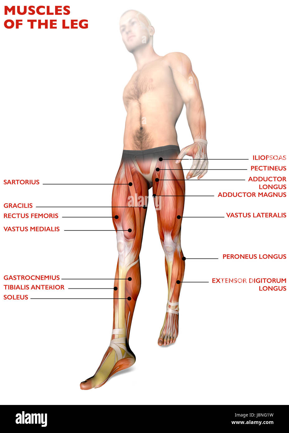
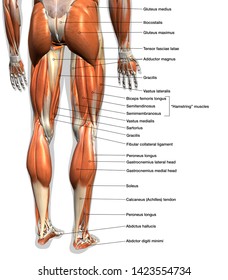
This sudden, tight, intense lower leg pain is sometimes called a charley horse. Direct impacts to the upper leg can damage the muscles and skin tissue, causing discoloration and pain. Take a look at the leg muscles diagram below, where you see each muscle clearly labeled. Supporting, balancing, and propelling the body is the work of the muscular system of the legs and feet. The human leg, in the general word sense, is the entire lower limb of the human. The short head originates from the lateral lip of linea aspera and. This group includes the adductor magnus, adductor longus, and adductor brevis muscles, as well as the pectineus and gracilis. A muscle located on the back portion of the lower leg, being one of the two major muscles that make up the calf:the flexing of this muscle during walking and bending of the knee creates traction on the femur, pulling it toward the tibia in the lower leg and causing the knee to bend. Collectively referred to as the hip adductors, the groin muscles are responsible for adduction of the hip, or drawing the leg in. The long head arises from a common tendon with semitendinosus from the superior medial quadrant of the posterior portion of the ischial tuberosity. The gastrocnemius muscle has two large bellies, called the medial head and the lateral head, and inserts into the calcaneus bone of the foot via its calcaneal tendon (also known as the achilles tendon.) The legs are the lower limbs of the human body that provide support and stability in addition to allowing movement. Related posts of muscle diagram leg muscle anatomy amazon.
Related posts of muscle diagram leg muscle anatomy amazon. The human leg, in the general word sense, is the entire lower limb of the human body, including the foot, thigh and even the hip or gluteal region. It is also visible on the medial edge of the thigh from the anterior. The groin muscles are a group of muscles situated high on the leg in the inner thigh. Notice the upper leg has a biceps muscle just like the upper arm does.
Leg Muscles Human Body Anatomy Muscle System 3d Rendering Stock Photo Alamy from c8.alamy.com The human leg, in the general word sense, is the entire lower limb of the human. This important tendon in the back of the calf and ankle stores the elastic energy needed for running, jumping, and other physical activity. The hamstring muscle attachment points. Illustration of human body anatomy from antique french art book: Related posts of muscle diagram leg muscle anatomy amazon. One of the most important tendons in terms of mobility of the leg is the achilles tendon. Collectively referred to as the hip adductors, the groin muscles are responsible for adduction of the hip, or drawing the leg in. Spend some time revising this diagram by connecting the name and location of each structure with what you've just learned in the video.
Pain in your calf or thigh can be caused by muscle cramps, a pulled or strained muscle, or issues related to your nerves.
The biceps femoris is a muscle of the posterior thigh composed of a long head and a short head. The legs are the lower limbs of the human body that provide support and stability in addition to allowing movement. Climbing stairs, standing, walking, and running are all activities that require strong contractions from the posterior muscle group to extend the leg. Collectively referred to as the hip adductors, the groin muscles are responsible for adduction of the hip, or drawing the leg in. Anterior compartment thigh muscles this is the largest of the three compartments of the thigh. The human leg, in the general word sense, is the entire lower limb of the human body, including the foot, thigh and even the hip or gluteal region. Notice the upper leg has a biceps muscle just like the upper arm does. Causes of upper leg pain related to trauma may include the following. See more ideas about muscle anatomy, human anatomy and physiology, body anatomy. These four muscles at the front of the thigh are the major extensors (help to extend the leg. The legs include the upper leg, knee, lower leg, ankle, and. The gastrocnemius muscle has two large bellies, called the medial head and the lateral head, and inserts into the calcaneus bone of the foot via its calcaneal tendon (also known as the achilles tendon.) Smooth muscles are found in the walls of many organs, such as the stomach and in blood vessels.
Www.purposegames.com *click them to make them larger & view details. One of the most important tendons in terms of mobility of the leg is the achilles tendon. Observe the leg muscle diagram posted above and notice that there are many parts in the muscles. Browse 435 leg muscle diagram stock photos and images available, or start a new search to explore more stock photos and images. Pain in your calf or thigh can be caused by muscle cramps, a pulled or strained muscle, or issues related to your nerves.
Leg Muscle Anatomy High Res Stock Images Shutterstock from image.shutterstock.com In the leg muscles diagram above, there are many muscles that make up your legs and support it to move. Muscle and bone anatomy 12 photos of the muscle and bone anatomy back muscles and bones anatomy, human muscle and bone anatomy, muscle & bone anatomy 3d free download, muscle and bone anatomy app, muscle and bone anatomy quiz, human muscles, back muscles and bones anatomy, human muscle and bone anatomy, muscle & bone. Spend some time revising this diagram by connecting the name and location of each structure with what you've just learned in the video. The muscles in the hip are responsible for the movement of the hip and, by proxy, the leg. Climbing stairs, standing, walking, and running are all activities that require strong contractions from the posterior muscle group to extend the leg. For women, shaping the thigh muscles is an essential goal of physical fitness. Direct impacts to the upper leg can damage the muscles and skin tissue, causing discoloration and pain. Muscle anatomy amazon 12 photos of the muscle anatomy amazon amazon muscle anatomy poster, muscle anatomy amazon, muscle anatomy model amazon, muscle trigger point anatomy amazon, human muscles, amazon muscle anatomy poster, muscle anatomy amazon, muscle anatomy model amazon, muscle trigger point anatomy amazon
One of the most important tendons in terms of mobility of the leg is the achilles tendon.
The muscles that make up the quadriceps are the strongest and leanest of all muscles in the body. From the large, strong muscles of the buttocks and legs to the tiny, fine muscles of the feet and toes, these muscles can exert tremendous power while constantly making small adjustments for balance — whether. Take a look at the leg muscles diagram below, where you see each muscle clearly labeled. The muscles work together to enable movement and keep the hip in alignment. On the medial edge of the posterior thigh is the gracilis muscle. See more ideas about muscle anatomy, human anatomy and physiology, body anatomy. The short head originates from the lateral lip of linea aspera and. These four muscles at the front of the thigh are the major extensors (help to extend the leg. Muscle and bone anatomy 12 photos of the muscle and bone anatomy back muscles and bones anatomy, human muscle and bone anatomy, muscle & bone anatomy 3d free download, muscle and bone anatomy app, muscle and bone anatomy quiz, human muscles, back muscles and bones anatomy, human muscle and bone anatomy, muscle & bone. Muscle of the human leg diagram in this image, you will find muscle of the human leg diagram, hip and femur middle layer, hip and femur deep layer, overview of the most important muscles of the leg, femur middle layer, femur deep layer, rectus femoris m. A muscle located on the back portion of the lower leg, being one of the two major muscles that make up the calf:the flexing of this muscle during walking and bending of the knee creates traction on the femur, pulling it toward the tibia in the lower leg and causing the knee to bend. Www.purposegames.com *click them to make them larger & view details. In the leg muscles diagram above, there are many muscles that make up your legs and support it to move.
/bodybuilder-working-leg-muscles-in-gym-1195621671-b74db540db4d47baab080f2ccf98d4a1.jpg)